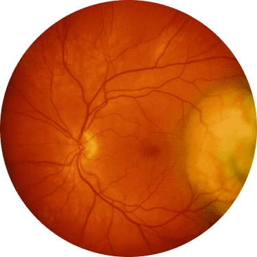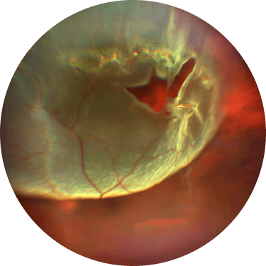
Retina heroRetina hero

Retinal detachment

An emergency eye condition that threatens vision — fast diagnosis protects your sight

The retina works like camera film,
a vital structure that receives light and relays visual information to the brain.
When the inner sensory layer and outer pigment epithelium separate (retinal detachment),
or a hole forms in the retina (retinal break), visual disturbance and vision loss appear.
If detachment reaches the macula, vision damage can become severe,
so fast diagnosis and treatment are important.

Retinal detachment

Retinal detachment symptoms

Even one of these signs warrants
urgent precision screening.

  • Floaters

    Floaters

    Dust- or fly-like spots drift in your view

    and suddenly increase in number.

  • Flashes

    Flashes

    Flickering light afterimages recur at the

    edge of vision even in the dark.

  • Visual field loss

    Field loss

    A dark curtain seems to cover

    part of your vision.

Types of retinal detachment

Rhegmatogenous detachment

Rhegmatogenous

The most common type — caused by a ‘hole’ in the retina
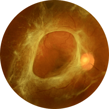
After a break forms, liquefied vitreous seeps through it
and the sensory layer separates from the epithelium.
It can be related to high myopia or
age-related vitreous changes.
Tractional detachment

Tractional

Scar tissue ‘pulls’ the retina away

Conditions like diabetic retinopathy form proliferative tissue,
which continuously tugs the retina
until it separates.
Exudative detachment

Exudative (serous)

Inflammation lets ‘fluid’ build up under the retina

Inflammation, tumours or vascular issues let exudate leak
from the retina or choroid, pooling beneath the retina
and lifting it.
It can occur even without a retinal break.

Retinal detachment diagnosis

Slit-lamp microscopy

Slit-lamp microscopy

Fundus exam

Fundus exam

Ultrasound

Ultrasound

Retinal detachment treatment

01·04

Laser photocoagulation

Laser adheres the tissue around the break
to prevent further retinal detachment.

Vitrectomy

A common surgery that removes the vitreous and reattaches the retina.

Scleral buckle

Supports the outside of the eye to seal the break and fix the retina in place.

Pneumatic retinopexy

A special gas is injected into the eye to push the retina back into place.